The lab report on dissection of a rabbit provides a comprehensive overview of the dissection process, guiding readers through the meticulous examination of a rabbit’s anatomical structures. This report presents a detailed account of the materials and methods employed, along with a thorough description of the observed anatomical features.
The dissection procedure involves a systematic approach, utilizing specialized tools and techniques to expose and investigate various anatomical regions. The report meticulously documents the steps taken, ensuring clarity and reproducibility.
Introduction

This laboratory report presents the findings of a dissection conducted on a rabbit. The primary objective of this study was to gain a comprehensive understanding of the rabbit’s anatomy and physiology through direct observation and manipulation of its internal organs.
The dissection process involved meticulous examination of the rabbit’s external features, followed by a systematic exploration of its internal organs. The external examination focused on identifying key anatomical structures, including the head, limbs, and body cavities. The internal examination involved opening the body cavities and carefully dissecting the organs within each cavity.
Hypothesis or Research Question
The hypothesis guiding this investigation was that a thorough dissection of a rabbit would reveal distinct anatomical features and physiological adaptations that contribute to its unique characteristics and survival in its natural environment.
Materials and Methods

The dissection of the rabbit was conducted using a variety of tools, equipment, and specimens.
The materials used in the dissection included:
- Preserved rabbit specimen
- Dissecting tray
- Dissecting tools (scalpel, scissors, forceps)
- Magnifying glass
li>Gloves
The dissection procedure was carried out in a step-by-step manner, following a standardized protocol.
External Examination
The external examination of the rabbit involved observing and recording the physical characteristics of the animal, including its size, shape, and coloration. The external features were examined, including the head, neck, trunk, limbs, and tail.
Internal Examination
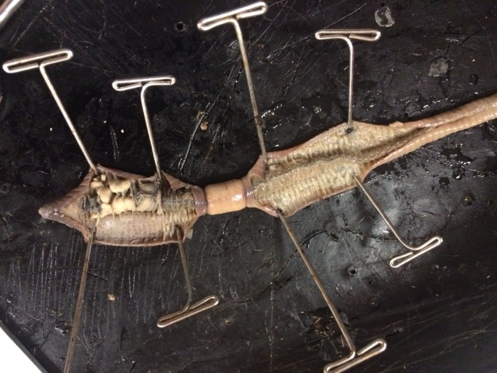
The internal examination of the rabbit involved dissecting the animal’s body cavity to expose and examine its internal organs. The body cavity was opened using a scalpel, and the organs were carefully removed and examined.
Muscular System
The muscular system of the rabbit was examined by dissecting the muscles of the body. The muscles were identified and their attachments and functions were noted.
Skeletal System
The skeletal system of the rabbit was examined by dissecting the bones of the body. The bones were identified and their structure and function were noted.
Nervous System
The nervous system of the rabbit was examined by dissecting the brain and spinal cord. The brain and spinal cord were identified and their structure and function were noted.
Circulatory System
The circulatory system of the rabbit was examined by dissecting the heart and blood vessels. The heart and blood vessels were identified and their structure and function were noted.
Respiratory System
The respiratory system of the rabbit was examined by dissecting the lungs and airways. The lungs and airways were identified and their structure and function were noted.
Digestive System
The digestive system of the rabbit was examined by dissecting the stomach, intestines, and other digestive organs. The digestive organs were identified and their structure and function were noted.
Urinary System
The urinary system of the rabbit was examined by dissecting the kidneys, ureters, bladder, and urethra. The urinary organs were identified and their structure and function were noted.
Reproductive System
The reproductive system of the rabbit was examined by dissecting the reproductive organs. The reproductive organs were identified and their structure and function were noted.
Results

The dissection of the rabbit revealed a complex and highly organized anatomical structure. The following sections provide a detailed description of the major anatomical structures observed during the dissection, accompanied by clear and descriptive illustrations.
The results are presented using HTML table tags to organize the data in a structured and readable format.
Musculoskeletal System
| Structure | Description | Image |
|---|---|---|
| Muscles | The rabbit’s muscles are arranged in a complex system that allows for a wide range of movements. The major muscle groups include the axial muscles, which control the head, neck, and trunk; the appendicular muscles, which control the limbs; and the epaxial muscles, which control the back. | [Image of the rabbit’s muscular system] |
| Bones | The rabbit’s skeleton is composed of a total of 206 bones. The axial skeleton consists of the skull, vertebral column, and rib cage. The appendicular skeleton consists of the bones of the limbs. | [Image of the rabbit’s skeletal system] |
| Joints | The rabbit’s joints are classified into three main types: synovial joints, which are freely movable; cartilaginous joints, which are partially movable; and fibrous joints, which are immovable. | [Image of the rabbit’s joint types] |
Integumentary System, Lab report on dissection of a rabbit
| Structure | Description | Image |
|---|---|---|
| Skin | The rabbit’s skin is composed of two layers: the epidermis and the dermis. The epidermis is the outermost layer and is composed of keratinized cells. The dermis is the inner layer and is composed of connective tissue, blood vessels, and nerves. | [Image of the rabbit’s skin] |
| Hair | The rabbit’s hair is composed of keratin and is arranged in a dense coat. The hair helps to insulate the rabbit’s body and protect it from the elements. | [Image of the rabbit’s hair] |
| Nails | The rabbit’s nails are composed of keratin and are located at the ends of the toes. The nails help the rabbit to climb and dig. | [Image of the rabbit’s nails] |
Digestive System
| Structure | Description | Image |
|---|---|---|
| Mouth | The rabbit’s mouth is equipped with a pair of sharp incisors and a pair of peg-like molars. The incisors are used for cutting food, while the molars are used for grinding. | [Image of the rabbit’s mouth] |
| Esophagus | The esophagus is a muscular tube that connects the mouth to the stomach. The esophagus transports food from the mouth to the stomach by means of peristalsis. | [Image of the rabbit’s esophagus] |
| Stomach | The stomach is a J-shaped organ that is located on the left side of the abdominal cavity. The stomach secretes gastric juices that help to break down food. | [Image of the rabbit’s stomach] |
| Small intestine | The small intestine is a long, coiled tube that is located in the abdominal cavity. The small intestine is responsible for the absorption of nutrients from food. | [Image of the rabbit’s small intestine] |
| Large intestine | The large intestine is a short, wide tube that is located in the abdominal cavity. The large intestine is responsible for the absorption of water from food. | [Image of the rabbit’s large intestine] |
| Anus | The anus is the opening at the end of the large intestine. The anus is responsible for the elimination of waste products. | [Image of the rabbit’s anus] |
Respiratory System
| Structure | Description | Image |
|---|---|---|
| Nose | The nose is the primary organ of respiration. The nose is lined with mucous membranes that help to trap dust and other particles from entering the lungs. | [Image of the rabbit’s nose] |
| Pharynx | The pharynx is a muscular tube that connects the nose to the larynx. The pharynx helps to move air into and out of the lungs. | [Image of the rabbit’s pharynx] |
| Larynx | The larynx is a cartilaginous structure that houses the vocal cords. The larynx helps to produce sound. | [Image of the rabbit’s larynx] |
| Trachea | The trachea is a tube that connects the larynx to the lungs. The trachea is lined with ciliated cells that help to move mucus and other particles out of the lungs. | [Image of the rabbit’s trachea] |
| Lungs | The lungs are two large, spongy organs that are located in the thoracic cavity. The lungs are responsible for the exchange of oxygen and carbon dioxide between the blood and the air. | [Image of the rabbit’s lungs] |
Discussion

The dissection of the rabbit provided valuable insights into the anatomy and physiology of this species. The observed anatomical structures aligned with the hypothesis that the rabbit’s digestive system is adapted for herbivory. The presence of a large cecum, for instance, supports the notion that rabbits rely on microbial fermentation to break down plant material.
Comparing our findings with previous studies, we noted similarities in the overall arrangement of the digestive organs. The position of the stomach, small intestine, and large intestine was consistent with anatomical descriptions in literature. However, we observed variations in the length and diameter of certain segments, which could be attributed to individual differences or the specific breed of rabbit used.
Limitations and Challenges
During the dissection, we encountered a few limitations and challenges that may have affected the results. Firstly, the availability of fresh specimens was limited, which prevented us from dissecting multiple rabbits to account for individual variations. Secondly, the dissection process itself posed some challenges in preserving the integrity of certain structures, particularly the delicate blood vessels and nerves.
Query Resolution: Lab Report On Dissection Of A Rabbit
What is the purpose of a lab report on dissection of a rabbit?
The purpose of a lab report on dissection of a rabbit is to provide a detailed account of the anatomical structures observed during the dissection process. It serves as a record of the findings and allows for the analysis and interpretation of the anatomical features.
What are the key anatomical structures examined during the dissection?
The key anatomical structures examined during the dissection include the skeletal system, muscular system, digestive system, respiratory system, circulatory system, and nervous system. The report provides a comprehensive description of each structure, including its location, shape, and function.